
Book a consultation

Home IDEL Diagnostic Programme Digital Infrared Thermal Imaging (DITI)

IDEL test 08 of 13

Thermography Digital Infrared Thermal Imaging (DITI)

Thermography, also known as thermal imaging, is an advanced diagnostic technique that utilizes infrared technology to detect and visualize temperature patterns and variations on the surface of the body.

warmercooler delta T
90%average sensitivity and specificity of breast thermography
800+peer-reviewed studies on breast thermography
1982FDA approval as an adjunctive breast cancer screening procedure
95%of early-stage cancers detected with a multimodal approach

01Understanding thermography

An innovative diagnostic tool for breast cancer and beyond

Thermography, also known as thermal imaging, is an advanced diagnostic technique that utilizes infrared technology to detect and visualize temperature patterns and variations on the surface of the body. This non-invasive and radiation-free imaging method has gained recognition for its potential diagnostic benefits, particularly in the detection and monitoring of breast cancer.

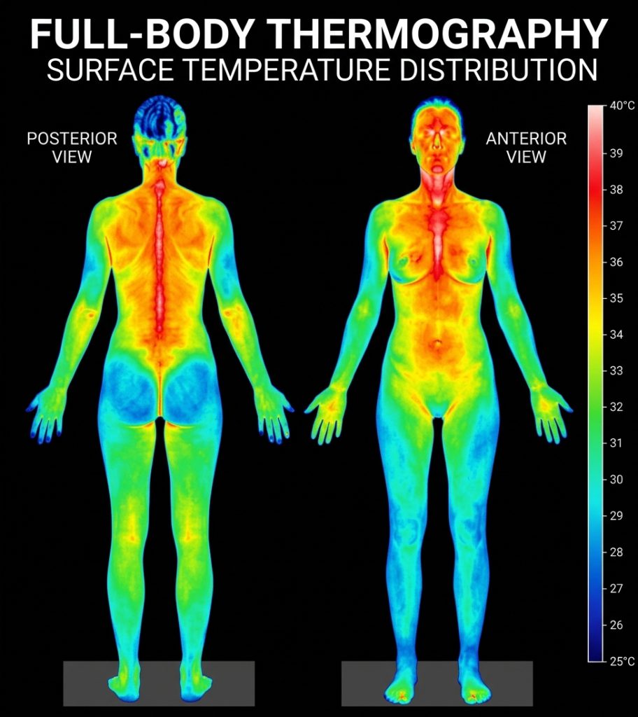
Thermography involves the use of specialized infrared cameras to capture thermal images of the body’s surface temperature distribution. These images display variations in heat patterns, highlighting areas of increased or decreased temperature. The underlying principle of thermography is that abnormal tissue activity, inflammation, and blood flow patterns can influence surface temperature, making it a valuable tool for detecting subtle changes that may be indicative of various health conditions.

A full-body thermal image from the back and the front, with a temperature scale from 25 to 40 °C
A full-body thermal image

02Diagnostic benefits for breast cancer

Five benefits for breast health

Early Detection

Thermography has the potential to detect early physiological changes associated with breast cancer. These changes, such as increased blood flow and abnormal temperature patterns, may occur before the development of detectable masses or structural abnormalities visible through other imaging modalities. Early detection increases the chances of successful treatment and improved outcomes.

Sensitivity and Specificity

Thermography demonstrates high sensitivity in detecting breast abnormalities. It can identify temperature asymmetry and subtle thermal patterns that may indicate abnormal tissue activity. Additionally, thermography has been shown to have a high negative predictive value, meaning that a normal thermogram can provide reassurance and reduce unnecessary further investigations.

Non-Invasive and Radiation-Free

Unlike mammography and other imaging techniques, thermography does not involve radiation exposure or physical compression of the breast. This makes it a safe and comfortable option, particularly for individuals who may be sensitive to radiation or discomfort during mammographic examinations.

Suitable for All Ages

Thermography is suitable for women of all ages, including those with dense breast tissue or breast implants. It can be used as an adjunct to other imaging modalities or as a standalone screening tool. Younger women with denser breasts often have a higher risk of false-negative results in mammography, making thermography a valuable complementary option.

Repeatable and Monitoring Capabilities

Thermography can be used for repeated screenings over time to monitor changes in temperature patterns. This enables healthcare professionals to track the progress of existing conditions, monitor treatment effectiveness, and detect any new or evolving abnormalities.

03Beyond breast cancer

Diagnostic benefits for other health problems

In addition to breast cancer detection, thermography holds diagnostic benefits for various other health problems, including:

Musculoskeletal Disorders

Thermography can assess inflammation, circulatory abnormalities, and nerve dysfunction associated with musculoskeletal conditions such as arthritis, sports injuries, and fibromyalgia. It aids in identifying areas of increased heat and can guide targeted treatment approaches.

Vascular Disorders

Thermography can help detect abnormalities in blood vessel functioning, assisting in the diagnosis and monitoring of conditions such as peripheral vascular disease, deep vein thrombosis, and diabetic neuropathy.

Inflammatory Conditions

Thermography can visualize temperature variations associated with inflammation, enabling early identification and monitoring of conditions like carpal tunnel syndrome, tendinitis, and bursitis.

Neuromuscular Disorders

Thermography can assist in diagnosing nerve entrapment, identifying regions of abnormal temperature, and guiding treatment planning for conditions such as carpal tunnel syndrome, sciatica, and neuropathies.

Detects hidden inflammation

Inflammation is the precursor to many cancers and other degenerative diseases such as arthritis, heart disease, stroke, diabetes and high blood pressure. Early detection of inflammation using Thermography may help prevent many negative health conditions from developing.

04Thermography can also

Function, not only structure

Tests of ‘anatomy’

X-rays, CT scans, Ultrasound, and MRI scans are all tests of ‘anatomy’ that are assessing the structure of the body

Thermography

thermography on the other hand is unique in that it measures physiological changes and functioning of the body, not only structural issues.

Visualize pain and pathology

Assess pain and pathology anywhere in the body

Is a very useful adjunctive procedure to other diagnostic tools, such as X-rays, bone scans, and ultrasound

Is very cost-effective, and risk-free as it uses no harmful radiation and provides you with instant images.

Fills in the gap in clinical diagnosis

05Other uses of thermography

Many other areas of health

Thermography can also be used in many other areas of health:

01

Early Detection of Heart Disease

Thermography also plays an important role in heart disease prevention as it can assess heart functioning and detect inflammation in the carotid arteries which may be a precursor to stroke and blood clots. Early detection of heart problems can save your life!

02

Arthritis

can help in its early detection and can differentiate between osteoarthritis and rheumatoid arthritis.

03

Neck and Back Problems

pain and joint degeneration appear very clearly on thermography scans and help to identify the source of the pain in the body

04

Dental Issues

thermography can also detect TMJ, gum disease, infected teeth, NICOs, and other dental issues that are precursors to many other serious diseases.

05

Sinusitis and Headaches

there is often a relationship between headaches and sinusitis – the thermography scan will show this very clearly.

06

Immune Dysfunction, Chronic Fatigue, and Fibromyalgia

when there is inflammation detected by thermography in the Thoracic spine areas of T1 and T2 this correlates well with immune system dysfunction.

07

Carpal Tunnel Syndrome

This is an often misdiagnosed condition – thermography can help to identify the source of the pain which may be from the cervical spine

08

Digestive Disorders

thermography can often see IBS, diverticulitis, and Crohn’s Disease and correlate these with other inflammatory issues in the body.

09

Other Health Issues

these would include bursitis, herniated discs, ligament and muscle tears, lupus, nerve problems, whiplash, cancer, and many others.

06Other interesting points about thermography

Decades of research

Joseph Bronzino mentions in his book entitled “Medical Devices and Systems” the following points about Thermography:

1982FDA approval

In 1982, the FDA approved breast thermography as an adjunctive breast cancer screening procedure.

1950sresearch since the late 1950s

Breast thermography has undergone extensive research since the late 1950s.

800+peer-reviewed studies

Over 30 years of research comprising over 800 peer-reviewed studies on breast thermography exist in the index-medicus literature.

300,000+women in the studies

In this database, well over 300,000 women have been included as study participants.

85Kparticipants in a single study

The numbers of participants in many studies are very large – 10K, 37K, 60K, 85K …

12 yearsof patient follow-up

Some of these studies have followed patients for up to 12 years.

15+ yearsof standardized protocols

Strict standardized interpretation protocols have been established for over 15 years.

90%sensitivity and specificity

Breast thermography has an average sensitivity and specificity of 90%.

10×more significant than family history

An abnormal thermogram is 10 times more significant as a future risk indicator for breast cancer than a first-order family history of the disease.

22×higher risk

A persistent abnormal thermogram carries with it a 22x higher risk of future breast cancer.

#1marker of high risk

An abnormal infrared image is the single most important marker of high risk for developing breast cancer.

10 yearsearlier than any other procedure

Breast thermography has the ability to detect the first signs that cancer may be forming up to 10 years before any other procedure can detect it.

61%better long-term survival

Research has shown that breast thermography significantly augments the long-term survival rates of its recipients by as much as 61%.

When used as part of a multimodal approach (clinical examination + mammography + thermography) 95% of early-stage cancers will be detected.

07How thermography works

Reading the heat of the skin

Thermography – is a non-invasive, diagnostic imaging procedure involving the detection and recording of cutaneous thermal patterns using instruments that can provide visual and quantitative documentation of these temperature measurements. Because thermography detects pathophysiologic changes in cutaneous temperature, it may be used as an integral part of the physical examination or, alternatively, as an adjunct to that examination.

Blood flow allows core temperature to come to the surface or not, and will change the temperature of the skin territorially. Each area of the body has a particular cutaneous-cordal connection by the sympathetic nervous system. The alteration of the thermal content of a territory occurs based upon alterations of this nervous control. Only the dermal blood flow changes explain the heat seen on the surface of the body.

The heat of a muscle, joint, or bone is not conducted to the dermal tissues and cannot influence the dermal temperature recorded by thermography. Conduction of heat from the deeper portions of the body to the surface does not occur or create changes in the surface temperature. However, there are neurochemical processes in the dermis that can block the normal sympathetic control of blood flow in the skin, such as histamines, prostaglandins, and myovascular receptor inhibitors of mast cells. These, however, can be easily differentiated from neural control by clinical correlation.

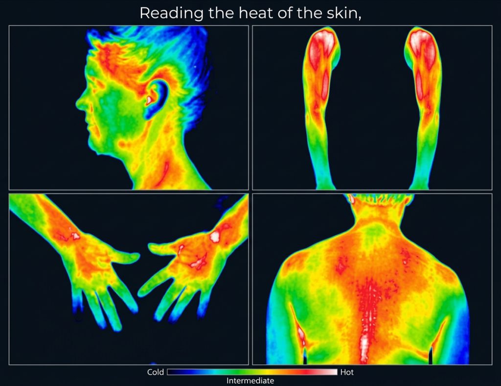
Thermal images of the head, arms, hands and back, on a scale from cold to hot
Thermal images of the head, arms, hands and back

Interpreting thermographic patterns

Symmetry and delta T

Areas of the surface of the body are recorded as symmetrical. The frontal areas, the thumbs, the great toes, the knees, the buttocks, and the posterior shoulders should be contralaterally symmetrical and isothermic. If there is a difference, it is described as a delta T and means simply the difference in temperature. In order to interpret thermographic patterns, one must know the normal pattern. The normal pattern of the skin surface is symmetrical, and it is thought that the 95 percentile range in the normal subject deviates less than 0.3° C. Uematsu and others have established the normal patterns of the human surface temperature.

Normal relationships

Relationships of various areas from one to another must be learned. The hands are usually colder than the shoulders. The buttocks and the feet are the coldest in the lower body, followed by the anterior knees. The thenar side of a given hand is usually warmer than the hypothenar, and the fingers are cooler than the palm.

Autonomic control

It has been shown that the surface temperature of the human body is symmetrical. It is a reflection of blood flow in the dermal microcirculation, and the control of this microcirculation is autonomic and specifically, sympathetic. The major basis of clinical thermography is the correlation of temperature recordings with various conditions from disease and injury as it relates to autonomic function.

Thermography is an innovative diagnostic tool that offers several advantages in the detection and monitoring of breast cancer and various other health conditions.

08Conclusion

A valuable complement to traditional imaging

Its non-invasive nature, early detection capabilities, high sensitivity, and suitability for all ages make it a valuable complement to traditional imaging techniques. By visualizing temperature patterns and variations, thermography provides healthcare professionals with important insights for timely intervention, personalized treatment planning, and ongoing monitoring. As research continues to advance, thermography holds promise for improving early detection rates, enhancing patient outcomes, and expanding its applications in the field of medical diagnostics.

DITI at the Da Vinci Centre

Digital Infrared Thermal Imaging (DITI) is practiced at the Da Vinci Holistic Health Centre in Larnaca. Images of the appropriate areas of the body are taken and sent to a team of specialist M.D. Thermologists in the USA. The images are analyzed and a full report is written and returned to the patient.

  1. Images taken

    of the appropriate areas of the body, in Larnaca

  2. Sent to the USA

    to a team of specialist M.D. Thermologists

  3. Full report

    written and returned to the patient

Dr. George Georgiou in his consulting room

Your practitioner

Dr. George Georgiou

Dr. Georgiou is a member of the American College of Clinical Thermology and a Certified Thermographer, a naturopath and has a Doctor of Science in Alternative Medicine.

Member, American College of Clinical ThermologyCertified ThermographerDoctor of Science in Alternative Medicine

↑↓ to chooseEnter to openEsc to close