Book a consultation

Home IDEL Diagnostic Programme Iridology

IDEL test 02 of 13

Iridology also known as iris analysis

Iridology, also known as iris analysis, is a unique diagnostic approach that examines the patterns and markings of the iris to gain insights into an individual’s health and potential disease predispositions.

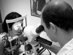
Dr. George examining a patient’s iris through the microscope Iris examination at the Da Vinci Centre
1880Peczely published a book describing principles of diagnostics using iris
5Central Hypotheses of Iridology, according to Dr. Bernard Jensen
28,000+iris nerve fibers, each controlling only 5 to 10 muscle cells
71.4–100%validity for predicting diseases, clinical trials at Aju University, South Korea

01Overview

Reading health in the coloured portion of the eye

The science of Iris Analysis or Iridology is an accurate and quick way of eliciting health information from the iris of the eye, the coloured portion. It is used by many health practitioners, including medical doctors in many countries throughout the world.

Iridology is a non-invasive practice that involves the examination of the iris, the colored portion of the eye, using a special microscope, for signs and markings that reflect the condition of various organs, tissues, and systems in the body. Iridologists study the structure, color, texture, and patterns of the iris to gather information about a person’s overall health and potential areas of imbalance or disease.

Marking in the iris can indicate inflammation in body tissues and organs, hereditary weaknesses, the constitution or our genetic ‘dowry,’ toxicity in the body, spinal subluxations, degeneration in organs and tissues, and much more.

Dr. George examining a patient’s iris through the microscope
Examination using a special microscope
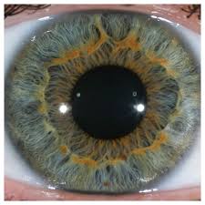
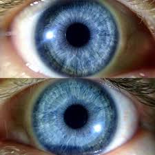
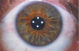
Close view of an iris, showing pigment and structure
Structure and colour
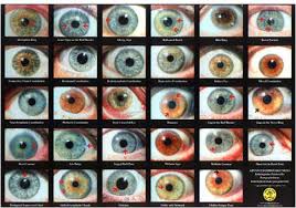
Iris constitutions
Iris constitutions

02What is looked for

Four kinds of markings

Iridologist usually looks for the following markings, as well as many more:

Color Variation

Different colors observed in the iris can indicate potential imbalances or predispositions. For example, dark or pigmented spots may suggest areas of increased toxicity or weakened organ function. Lighter spots may indicate potential weaknesses or areas of inflammation.

Structural Patterns

Iridology recognizes various structural patterns in the iris, such as crypts, furrows, and lacunae. These patterns may suggest potential weaknesses or imbalances in specific organs or systems. For instance, a prominent crypt in a certain area may indicate susceptibility to digestive issues.

Radial and Concentric Patterns

The iris may exhibit radial and concentric patterns, which iridologists interpret as indicators of lymphatic congestion, toxicity, or inflammation in specific areas of the body. These patterns can provide insights into potential underlying health issues that need attention.

Pupillary Border

The pupillary border of the iris is another area of interest in iridology. Distinct markings, such as rings or discoloration, may indicate potential weaknesses or imbalances in specific organs or body systems. For instance, a dark ring around the pupil may suggest potential kidney or adrenal gland dysfunction.

03History

How iridology began

  1. 1826 – 1907

    Ignatz von Peczely

    Egernar, near Budapest, Hungary

    The medical doctor named Ignatz von Peczely (1826-1907) from Egernar, near Budapest, Hungary has proved the basis of the method and proposed his own iris zones projection chart although rather primitive from modern point of view but relatively reliable. The scientist’s biographers give rather interesting legend determined the mission of the future founder of modern iridology.

  2. 1880

    The first iris chart

    Viennese University

    While studying in the Viennese University and working in the surgery hospital, Peczely started to investigate the patient’s iris changes depending on different diseases. He has found that each part of the body as well as each organ correspond to the determined iris segment. As a result the first iris chart was developed making him a founder of modern iridology. In 1880 Peczely published a book describing principles of diagnostics using iris. The main attention of his work was the location and form of iridological signs. Later he has published a manual on iris diagnostics where he wrote as epigraph that eyes are not only mirroring of the soul but of the body as well. This work was met by icy silence of contemporaries.

  3. Sweden

    N. Liljequist

    Swedish pastor

    Another important page to iridology history was added by Swedish pastor N. Liljequist. ‘A man can lie but his eyes never lie’ was his answer to those who were interested in the reasons of his interest to the iridology. He developed a more detailed iridology chart, working independently of Peczely that was more reliable and better.

A man can lie but his eyes never lie
N. Liljequist, Swedish pastor

04Basis of iridology

The five central hypotheses

According to Dr. Bernard Jensen, the Central Hypotheses of Iridology are:

1Hypothesis

Pigment and structure

The iris reveals, through changes in pigment and structure, abnormal conditions of tissue in the human body.

Close view of an iris, showing pigment and structure
Pigment and structure, as seen in the iris
2Hypothesis

Topography

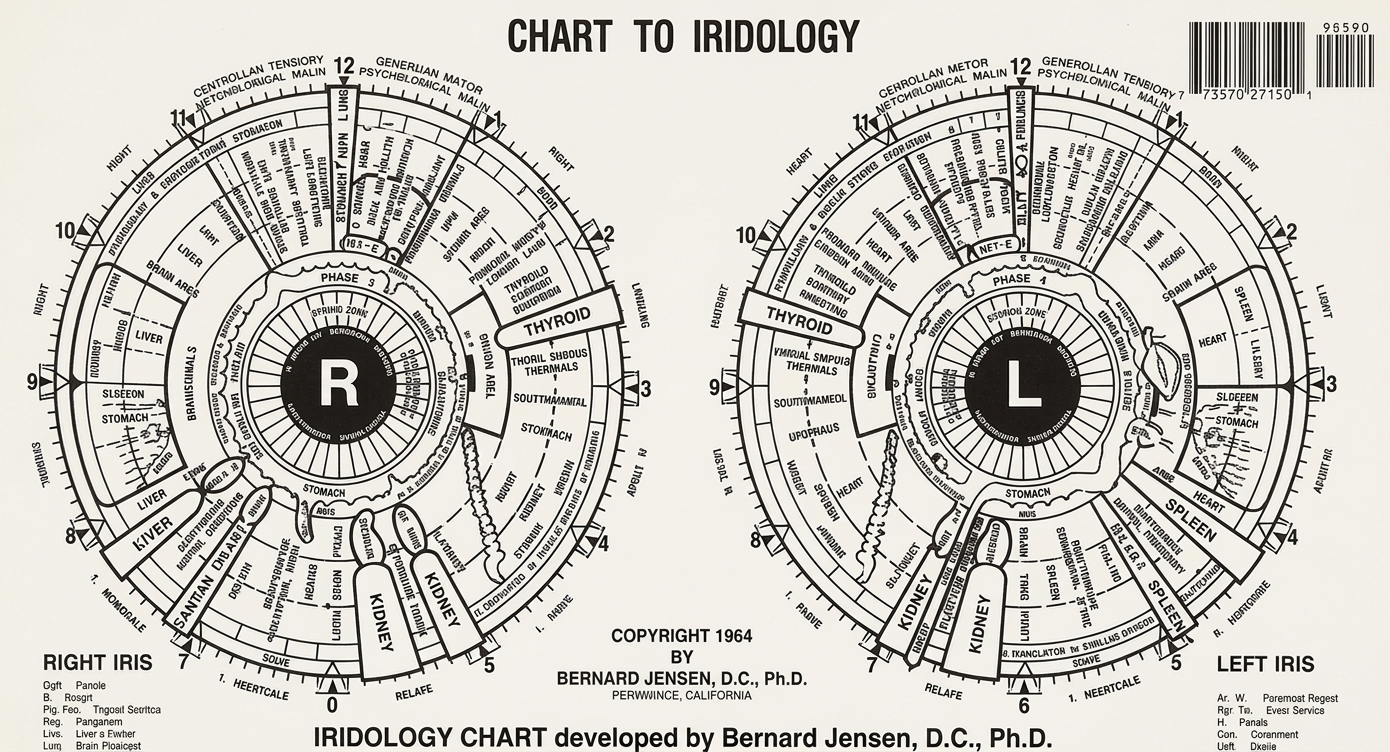
The anterior of the iris reflexly corresponds, in the systematic organization of its topography, to the major tissue structures the body. For example, each organ, gland and tissue is represented in a specific location in the left or right iris, or both. The chart indicated below is self-explanatory.

Iridology chart developed by Bernard Jensen, D.C., Ph.D., showing the right and left iris
Iridology chart developed by Bernard Jensen, D.C., Ph.D.
3Hypothesis

Left and right

Organs and tissues on the left half of the body are reflexly represented in the left iris, while those of the right half of the body are represented in the right iris. Organs and tissues lying along the centreline of the body, the sagittal plane, appear in both irides, as do bilateral organs.

Iris5 to 10
Extremities200 – 300

Muscle cells controlled by a single nerve fiber. This means that changes reflexed to the iris are highly specific to certain zones.

A pair of irides
Both irides

The neural pathway proposed by Walter Lang

  1. Organs and tissues of the bodyafferent autonomic nerve impulses
  2. Anterior thalamic nuclei of the diencephalona “file cabinet” or storage area
  3. Hypothalamusa central relay and control station of the brain
  4. Oculomotor nucleus and the Edinger-Westphal nucleus
  5. Muscles, stroma and blood vessels of the iridestopographical tissue changes
The research behind this hypothesis

Again, this hypothesis is based upon the findings and empirical observations of hundreds of Iridologists. The research of Walter Lang (Die anatomischen und physiologischen Grundlagen der Augendiagnostik) proposes a set of neural pathways that would account for the physiological eye changes associated with both disease states and abnormal tissue conditions. Lang indicates that afferent autonomic nerve impulses from the various organs and tissues of the body reach the anterior thalamic nuclei of the diencephalon. It therefore acts as a “file cabinet” or storage area on all anatomical conditions. Lang further suggests that this information is transmitted to the hypothalamus, which functions as a central relay and control station of the brain.

Nerve impulses from the hypothalamus follow pathways via the oculomotor nucleus and the Edinger-Westphal nucleus to the muscles, stroma and blood vessels of the irides, where they stimulate topographical tissue changes, corresponding to specific organ and tissue changes elsewhere in the body. Lang shows that areas of the iris can be differentiated to correspond to the various organs and tissues by definition that a single iris nerve fiber (over 28,000) controls only 5 to 10 muscle cells as compared to 200 – 300 muscle cells in the extremities. This means that changes reflexed to the iris are highly specific to certain zones. However the neural system organization is the same in all human beings, the same tissue areas of the body will always reflex to the same areas of the irides.

4Hypothesis

The anterior iris

The anterior iris, including the anterior epithelium, the stroma, the muscle layer, the pupillary margin, the autonomic nerve wreath (collarette), and the scleral-iris margin undergo specific changes corresponding to pathological changes in specific organs and tissues in the body.

How the change shows in the iris fibers

WhitenessacuteSub-acuteChronicDegenerativeincreasing darkness
12345
  1. 1The pupillary margin
  2. 2The autonomic nerve wreath (collarette)
  3. 3The stroma
  4. 4The ciliary zone · Hypothesis 5
  5. 5The scleral-iris margin
The research behind this hypothesis

Again, this hypothesis is based upon findings and empirical observations of hundreds of Iridologists. Iridologists have distinguished that the iris changes due to pathological deterioration of body tissue. This shows as a whiteness (acute) in the iris fibers, then an increasing darkness (sub-acute -> chronic -> degenerative) and the depth of iris lesions. In the past, Iridologists have also stated that they have witnessed changes due to reversal pathology (emergence of healing lines.) in the same iris lesions after successful treatment. In many cases these findings are confirmed with other commonly accepted diagnostic procedures.

It must be emphasized that the pathological condition must be adequately severe to be classified by the standards of western medicine as the classification standards of Iridology are different. The various laboratory tests that are being used to determine the presence or absence of disease are not totally efficient in the assessment of sub-clinical conditions. Acute and sub-acute stages of tissue inflammation can commonly be detected long before other diagnostic methods are capable of finding any health problems with the patient. In a similar manner, when a patient is declared “well” by the standards of Western medicine, Iridology frequently shows a sub-acute condition persisting in the individual.

German medical researcher, Walter Lang, has demonstrated that the autonomic nerve fibers from virtually every gland, organ and tissue of the body extend to the thalamus and hypothalamus which monitor and respond to changes of condition in all anatomical structures. These changes of state, Lang suggests, are relayed from the thalamus and hypothalamus through the ophthalmic branch of the trigeminal ganglion to the motorneurons of the iris muscle structure.

Changes in the impulses conducted by these motorneurons may be responsible for the changes in the muscle structure of the iris, leading to the gradual separation of iris fibers in the stroma and consequent appearance of the lesions and other markings familiar to iridologist. Lang also points out that the organization of the human nervous system is genotypic, and further postulates that innervation to the iris reliably represents is also genotypic, which accounts to the fact that the iris reliable represents the same organs, glands, and other anatomical subdivisions of the body in precisely the same locations in the irides of all individuals.


5Hypothesis

Strengths and weaknesses

Inherent weaknesses, inherent strengths and the degree of nervous system sensitivity are shown in the iris, respectively, by the crypts and separations in the trabeculae (the fibres in the iris); by closely knit trabeculae; and by parallel, curved cramp rings concentric with outer perimeter of the iris, all located in the ciliary zone outside the autonomic nerve wreath.

Inherent weaknessesthe crypts and separations in the trabeculae (the fibres in the iris)
Inherent strengthsclosely knit trabeculae
The degree of nervous system sensitivityparallel, curved cramp rings concentric with outer perimeter of the iris
all located in the ciliary zone outside the autonomic nerve wreath
Iris showing crypts and trabeculae
Crypts and trabeculae in the iris
The research behind this hypothesis

Scientific research has demonstrated that the posterior pigment epithelium and dilator muscle of the irides are embryologically derived from neurectoderm, the tissue from which the central nervous system (brain and spinal cord) are also derived. Iridologist find that it is this similarity that reflects in the iris the genetic inheritance of the individual. It is assumed that the specific configuration and development of the dilator muscle somehow determine the radial arrangement and spacing of the vascular arcades (trabeculae) in the stroma above it. The fact that crypts and separated trabeculae represent inherent weaknesses in no way conflicts with the fact that the same structures allow aqueous humor to circulate through the irides.

05Accuracy

What is the accuracy of iris analysis?

The accuracy and reliability of iris signs as reflex indications of tissue pathology in the body have been confirmed in many thousands of instances by: laboratory tests, X-rays, and other commonly accepted diagnostic approaches. Recent research from clinical trials in South Korea from Aju University have shown that the validity of using iridology for predicting diseases ranges between 71.4% and 100% accuracy.

71.4% – 100%Validity of using iridology for predicting diseases — clinical trials, Aju University, South Korea

The accepted clinical results by the Korean Government include:

Diseases of: % Accuracy

The accepted clinical results by the Korean Government include: Diseases of: % Accuracy
Digestive system90.2%
Endocrine system86.4%
Urogenital system85.7%
Circulatory system81.6%
Nervous system79.9%
Cardiovascular system75%
Muscle/Skeletal system72.2%
Immune system54.2%
Dr. George Georgiou

Your iridologist

Dr. George Georgiou

Dr. Georgiou is a qualified Iridologist and a member of the Guild of Naturopathic Iridologists, UK. He is also an active member of the Canadian Neuro-Optic Research Institute, Canada where he works closely with Prof. Bryan Marcia, its founder and holder of a Ph.D. in Iridology.

Guild of Naturopathic Iridologists, UKCanadian Neuro-Optic Research Institute, Canada

Anyone interested in studying this fascinating subject go HERE.

↑↓ to chooseEnter to openEsc to close